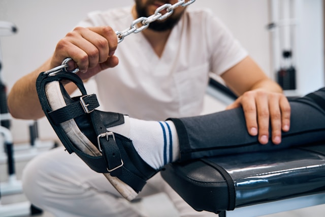
Orthopedic surgery has transformed into a discipline where Precision Orthopedic Techniques guide every decision, from preoperative planning to postoperative recovery. Surgeons deliver safer, more effective treatments by leveraging detailed imaging, computer navigation, robotic assistance, minimally invasive strategies, biologic enhancements, and personalized rehabilitation. This article explores five critical areas—foundations, imaging and navigation, robotics and power tools, minimally invasive and biologic approaches, and patient-centered rehabilitation—demonstrating how Precision Orthopedic Techniques improve outcomes and pave the way for future innovations.
Precision Orthopedic Techniques: Foundations of Accurate Fracture Repair
The roots of Precision Orthopedic Techniques date to the late 19th century, when Dr. Hugh Owen Thomas introduced the plaster cast. Before that, fracture management varied widely, often resulting in poor alignment and long recoveries. The standardized use of casts allowed surgeons to maintain bone position more consistently, reducing malunion and enhancing functional outcomes.
World War I spurred further advances. Surgeons like Sir Robert Jones pioneered internal fixation by inserting metal plates and screws directly into fractured bones. This enabled earlier mobilization, minimizing muscle atrophy and joint stiffness. Although early implants lacked today’s specialized alloys, they embodied the core principle of Precision Orthopedic Techniques: creating a stable mechanical environment that supports biological healing.
By the mid-20th century, improved sterilization and antibiotics made complex reconstructions safer. Surgeons began correlating intraoperative observations with preoperative X-rays, refining reduction techniques. These foundational innovations proved that meticulous planning and tailored hardware are essential for precise fracture care, principles that underpin modern Precision Orthopedic Techniques.
Precision Orthopedic Techniques: Innovations in Imaging and Navigation
Real-time and three-dimensional imaging have redefined surgical precision. The introduction of fluoroscopy in the mid-20th century allowed intraoperative X-ray guidance so surgeons could instantly confirm hardware placement. For instance, in distal radius fractures, fluoroscopy ensures screws avoid joint surfaces, preventing arthritis.
Computed tomography (CT) scanners later provided detailed 3D models of bone and soft tissues. Complex pelvic and acetabular fractures—once navigated through a combination of skill and X-ray interpretation—became manageable through CT-based planning. Surgeons could visualize fragment orientation, plan screw trajectories, and anticipate challenges before making an incision.
Building on these imaging advances, computer-assisted navigation systems emerged in the 1990s. Navigation platforms guide tools with sub-millimeter accuracy by fusing CT or MRI data with optical trackers on instruments. In spinal fusion, navigation ensures pedicle screws follow the planned corridor, reducing neural injury risks. Such systems make Precision Orthopedic Techniques predictable, reproducible, and safer.
Precision Orthopedic Techniques: Robotics and Power-Assisted Tools
Beyond navigation, powerful instruments and robotics have enhanced both speed and precision. High-speed burrs, oscillating saws, and ultrasonic osteotomes allow clean, controlled bone resections while preserving soft tissue. An ultrasonic osteotome can create bone tunnels with exact dimensions in anterior cruciate ligament reconstruction, improving graft fit and reducing failure rates.
Robotic assistance represents the pinnacle of Precision Orthopedic Techniques. Systems like the MAKO™ robot for knee arthroplasty and ROSA™ for spinal alignment translate surgeon-designed plans into highly accurate movements. Haptic feedback prevents deviation from planned resection boundaries, while real-time tracking confirms tool position. Studies show robotically assisted knee replacements achieve mechanical alignment within one to two degrees of the ideal axis, which correlates with longer implant survival and better functional recovery.
These robotic platforms also collect intraoperative data, feeding back into surgical planning algorithms. As more procedures use robotic assistance, the knowledge of optimal techniques grows, continually refining precision orthopedic techniques and improving patient outcomes.
Precision Orthopedic Techniques: Minimally Invasive and Biologic Strategies
Patient demand for smaller scars and faster recovery has driven the spread of minimally invasive methods under Precision Orthopedic Techniques. Arthroscopy—using tiny portals and a fiber-optic camera—allows meniscal repairs, cartilage debridement, and ligament reconstructions with minimal soft-tissue disruption. Compared to open surgery, patients experience less pain, shorter hospital stays, and quicker return to activities.
Percutaneous fixation extends these principles to fracture care. Under fluoroscopic or navigation guidance, surgeons insert intramedullary nails and screws through small incisions, aligning fractures within the bone’s medullary canal. Early weight-bearing is often possible, reducing rehabilitation time.
Biologic enhancements now complement mechanical repairs. Platelet-rich plasma (PRP) concentrates growth factors from the patient’s blood to promote tendon and ligament healing. PRP injections around the repair site have increased tendon-to-bone integration in rotator cuff surgery. Bone morphogenetic proteins (BMPs) stimulate new bone formation in spinal fusions, reducing the need for bone graft harvest. Tissue-engineered scaffolds loaded with stem cells show promise for regenerating cartilage defects. By integrating biologic agents into workflows guided by Precision Orthopedic Techniques, surgeons optimize the mechanical and biological aspects of healing.
Precision Orthopedic Techniques: Personalized Rehabilitation for Optimal Outcomes
True precision extends into postoperative rehabilitation. Wearable sensors—such as inertial measurement units and pressure-sensing insoles—capture data on joint angles, loading patterns, and gait symmetry. After a total hip replacement, sensors in the patient’s shoe can detect subtle limp patterns. Physical therapists then tailor exercises to correct these deviations, preventing compensatory injuries.
Virtual reality (VR) rehabilitation adds an immersive dimension. Patients recovering from ankle fractures navigate a simulated trail, with software providing visual feedback on balance and range of motion. This gamified approach increases engagement and adherence to rehab protocols.
Telemedicine platforms integrate these data streams, allowing surgeons and therapists to monitor progress remotely. If a patient’s knee flexion plateaus, the care team can adjust the exercise regimen in real time. This feedback loop ensures Precision Orthopedic Techniques delivers lasting benefits beyond the operating room.
By combining historical insights with imaging, navigation, robotics, minimally invasive and biologic methods, and personalized rehabilitation, Precision Orthopedic Techniques has redefined musculoskeletal care. As technologies such as additive manufacturing, artificial intelligence, and smart implants mature, the future promises ever-greater accuracy, enhanced power, and individualized treatment plans, ensuring that each patient receives the best possible outcome.